Updates on intraoperative MRI applications in neurosurgery
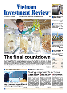
 |
| Hitachi's new iMRI scanner offers an untold number of advantages for Vietnamese neurosurgeons |
On this occasion, the Intraoperative MRI (iMRI) was also introduced for the first time, expecting to foster a leap in neurosurgery in Vietnam.
The workshop attracted the attendance of a great number of leading experts and doctors from Viet Duc Hospital, K Hospital, Bach Mai Hospital, Cho Ray Hospital, Vinmec Hospital, International Neurological Hospital, and Friendship Hospital, among others. The experts and doctors have exchanged useful knowledge and information.
Especially Assoc. Prof. Dr. Dong Van He, director of Viet Duc Hospital's Centre for Neurosurgery and Dr. Yoshihiro Muragaki from Department of Neurosurgery at Tokyo Women Medical University brought updated information about iMRI applications in neurosurgery.
“Performing a brain surgery is like driving a race car. The timing of each pit stop is critical for success. As such, iMRI was invented to provide a solution for faster and safer MRI scanning during surgery,” said Dr. Muragaki.
 |
| Dr. Muragaki updating participants on the advantages of iMRI scanning |
Neurosurgery is one of the most difficult and complex surgical procedures because cerebrovascular interventions require millimeter precision to minimise affects to the surrounding normal tissues as well as limit blood loss, pain, and risk of complications during surgery.
Prior to surgery, the patient will receive an MRI examination to determine the location and dimensions of the tumor. However, after the opening of the skull, these tumors are displaced by increased intracranial pressure, leading to difficulties for the naked eye to accurately determine the tumor’s boundaries.
After the surgery, the patient will have another MRI examination to determine whether the tumor was removed completely, and in some cases, resuscitation is needed to remove the residual lesion after the first surgery.
Thanks to science and technology advancement, iMRI technology is now available in Vietnam, enabling physicians to fully address these issues.
iMRI - One room solution
In some countries with developed healthcare systems, MRI solutions have been applied in two models: two-room and one-room design.
Two-room design (two separate rooms for MRI and operation) has some disadvantages, such as increased risk (due to lengthy patient-shifting intervals between MRI imaging and surgery), and costs (due to space constraints). Meanwhile, time is a decisive factor in the success of neurosurgery.
To address these disadvantages, Hitachi provides an intraoperative solution with its “medium/low field open one-room MRI system,” where the MRI system is in the operating room, allowing surgeons to perform MRI scans whilst the surgery is on-going. Surgeons need not worry about the interference between the magnetic field and the surgical equipment or the MRI display because they are compatible with the magnetic field.
 |
| The event saw the participation of leading experts and doctors from Vietnamese medical institutes |
With Hitachi’s one-room solution, the interval between MRI imaging and surgery is minimised by eliminating the need to roll the patient from the operating room to the MRI and back again.
Furthermore, Hitachi’s operating table is designed with a rotating mechanism, which allows surgeons to move the patient over to the MRI cabinet safely in one swift motion. All of this serves to minimise the time between MRI imaging and surgical procedures, for a safe and effective surgery for patients.
MRI scan in the middle of surgery
Being able to perform MRI scans on the patient whilst the surgery is on-going provides surgeons with real time information which can greatly aid decision-making in the operating room. Given this feature, surgeons can use real time iMRI scans to verify surgical results and detect any remaining tumors before the surgery is over.
This allows surgeons to perform immediate rectification and achieve complete resection in a single surgery. With iMRI support, surgeons can maximise their potential and achieve better surgical outcomes for patients.
Besides, Hitachi’s one room system uses an open MRI design with which surgeons can easily observe the patient, reducing surgical risks. Furthermore, high-sensitivity solenoid receiver coils can be installed directly on the fixed head frame, further boosting MRI imaging precision. All above features can be installed and used together with existing surgical instruments and equipment, which helps save costs for hospitals.
“Thanks to advanced applications in operation, especially neurosurgery, physicians will be better supported in the operating room, which helps minimise risks that may occur during operation, allowing patients to access better healthcare services,” said Assoc. Prof. Dr. Dong Van He, director of Viet Duc Hopital’s Centre for Neurosurgery.
 |
| Prof. Dr. Dong Van He speaking at the event |
Ngo Thanh Son, general director of JVC, said: "Neurosurgery remains a concern of medicine and requires absolute speed and accuracy. This is the driving force for us to bring the best products and services that help patients and families feel secure during surgery.”
Japan Vietnam Medical Instrument Company (JVC) was established on September 27, 2001. After 15 years of construction and development, JVC has supplied and installed thousands of modern medical systems, especially ones used in imaging diagnostics, such as Magnetic Resonance Imaging System, Computed Tomography System, X-ray System, X-ray digitisation system, and ultrasound system, among others.
The company also provides the best technical services to ensure the stable operation of these systems for the purposes of supporting hospitals, medical centres, and clinics in providing advanced medical services for patients.
What the stars mean:
★ Poor ★ ★ Promising ★★★ Good ★★★★ Very good ★★★★★ Exceptional
Latest News


More News
- The generics industry: unlocking new growth drivers (February 04, 2026 | 17:39)
- Vietnam ready to increase purchases of US goods (February 04, 2026 | 15:55)
- Steel industry faces challenges in 2026 (February 03, 2026 | 17:20)
- State corporations poised to drive 2026 growth (February 03, 2026 | 13:58)
- Why high-tech talent will define Vietnam’s growth (February 02, 2026 | 10:47)
- FMCG resilience amid varying storms (February 02, 2026 | 10:00)
- Customs reforms strengthen business confidence, support trade growth (February 01, 2026 | 08:20)
- Vietnam and US to launch sixth trade negotiation round (January 30, 2026 | 15:19)
- Digital publishing emerges as key growth driver in Vietnam (January 30, 2026 | 10:59)
- EVN signs key contract for Tri An hydropower expansion (January 30, 2026 | 10:57)


 Mobile Version